
by Miriam Valera-Alberni
Mitochondria: the tiny energy producers of the cell
Every human is made of an estimated 37 million cells, each of which contain compartments known as ‘organelles’, specific subunits with unique functions. While the nucleus provides the genetic instructions to the cell, mitochondria are key to bioenergetics and metabolism of the organism. Even though they are essential to keep us alive, mitochondria were not always part of our cells.
| When life was non-existent, oxygen levels were nearly zero. The presence and expansion of oxygen-producing organisms (such as plants) influenced in the increase of oxygen levels, which threatened the proto-eukaryotic cell that lived in no-oxygen conditions. However, the symbiosis of an ancient proteobacteria (the ancestor of mitochondria) – which was able to use oxygen – by the proto-eukaryotic cell contributed not only to its survival, but also to the evolution of eukaryotic cells and more complex forms of life (Figure 1). This establishes, therefore, an example of how environmental change triggers evolutionary events. Figure 1. Endosymbiotic theory. Symbiosis refers to the relationship between two species that benefit from living and working together. When one of the species actually lives inside the other it’s called endosymbiosis. The endosymbiotic theory describes how the ancient proto-eukaryotic cell ingested a proteobacteria, resulting in a permanent collaboration that would lead to today’s eukaryotic cell. Source: University of Utah (The Evolution of the Cell). | 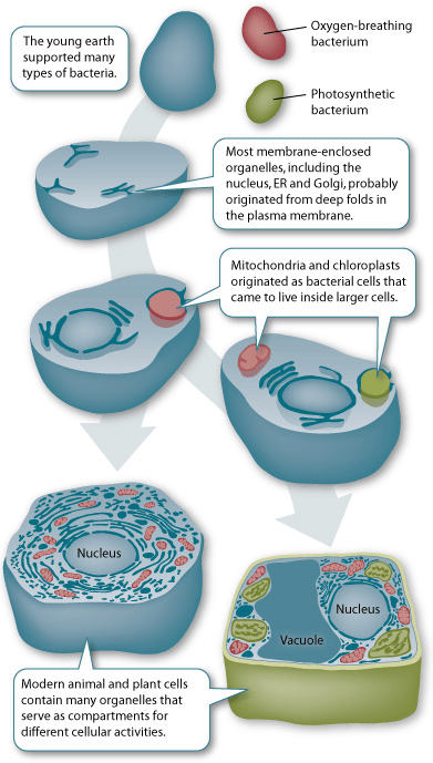
|
Mitochondria use oxygen to process the nutrients that arrive to our cells, and in return, the organelle produces the chemical energy (ATP) that the cell – and the organism – needs to carry on living. This is the reason why mitochondria are widely known as the ‘powerhouses of the cell’. Nevertheless, far from being their only role, mitochondria also act as quality control checkpoints, participating in a wide range of cellular events beyond energy production. These include the regulation of cell growth, cell division, calcium homeostasis, lipogenesis and the production of steroid hormones. Mitochondria can even determine cell fate, triggering signaling cascades leading to cell death. The origins of mitochondria also explain how they have their own DNA (mitochondrial DNA or mtDNA), which codes only for 13 mitochondrial proteins, while the rest of the proteins that mitochondria need (approximately 1500 in mammals) are encoded by the nucleus (nuclear DNA or nDNA). Thus, the mitochondria and the nucleus act as two independent entities which communicate continuously.
Mitochondria as a dynamic network
Mitochondria were originally conceived as solitary and static entities in the cytoplasm. However, far from that, the mitochondrial network is highly dynamic and adapts in size and morphology to changes in the cellular environment (Figure 2). For instance, starvation leads to the fusion of mitochondria, which increases the oligomerization of ATPase subunits. This maintains ATP production and contributes to the survival of the starving cells. On the other hand, fragmented mitochondria were observed in diet-induced and genetic obese mouse models, associated with mitochondrial dysfunction and insulin resistance. Therefore, changes in mitochondrial morphology correlate with various cellular and environmental stresses, and this alteration in mitochondrial shape contributes to their final function.


Figure 2. On the left, old perception of the cell, in which mitochondria are depicted as individual bean-shaped entities. However, visualizing live cells under the microscope reveals mitochondria (on the right, in green) in different shapes, located closely to the nucleus and extending throughout the cell.
The mitochondrial life cycle includes events of mitochondrial content increase (biogenesis), removal of damaged mitochondria (mitophagy), or rearrangement of the mitochondrial network by fusion/fission (mitochondrial dynamics). Mitochondrial dynamics refers to the repetitive and coordinated cycles of fusion (two or more mitochondria come close in contact and can fuse their membranes) and fission (one mitochondrion divides into two daughter mitochondria). To have an idea of how mitochondrial dynamics looks like, click in the following link here. Mitochondrial dynamics impacts on mitochondrial function, as the rate of fusion/fission contributes to the balance between health and disease. Mutations in the key players of mitochondrial dynamics have been associated with numerous human pathologies, including degenerative disorders (Parkinson’s or Alzheimer’s), neuromuscular disorders (Charcot–Marie–Tooth disease) and metabolic disorders (diabetes and obesity).
Mitochondria inheritance
The conventional conception is that mitochondria are only inherited from the mother, because when sperm fertilize an egg, only the nucleus’ genetic information pass on to the embryo, while the rest (including the father’s mitochondria) is lost while entering the egg (Figure 3). Therefore, the embryo has all the mitochondria from the mother’s egg. However, researchers have recently identified few cases in which the mitochondrial DNA (mtDNA) could be inherited from both parents. As mtDNA sequencing is only performed when a mitochondrial disease is suspected, it is not clear how frequent this event might take place or whether the bi-parental inheritance is associated to pathogenicity or mitochondrial dysfunction.
 Figure 3. Conventional mtDNA inheritance. Unlike nuclear DNA, which is inherited half from mother and half from father, mitochondrial DNA is passed only from females. When the sperm fertilizes the egg, it leaves behind all its mitochondria: the embryo then only contains maternal mitochondria. Source: “Inheritance of mitochondrial and chloroplast DNA”, Khan Academy (public domain).
Mitochondrial diseases
Because mitochondria are critical in cell life, mitochondrial dysfunction results in an array of organ malfunctions and diseases. Mitochondrial diseases are the result of either inherited or spontaneous mutations in the mitochondrial DNA, but can also appear from mutations in the nuclear-encoded mitochondrial proteins.
Mitochondrial diseases target parts of the body that require the greatest amounts of energy, such as the heart, brain and muscle. While some mitochondrial disorders only affect a single organ (i.e, the eye in LHON syndrome – Leber hereditary optic neuropathy), others involve multiple organ systems and present a more complicated symptomatology (such as the Leigh syndrome, characterized by progressive loss of mental and movement abilities). Raising awareness is essential to educate people about mitochondrial diseases, but also provides a time for the affected families to come together and get in contact with the research carried out in the laboratories (Figure 4). Establishing a proper diagnosis can be relatively straightforward when a patient has a recognizable phenotype, which is supported by the family’s history and validated by molecular genetic testing.
Overall, the involvement of mitochondria in the health/disease balance emphasizes the importance of understanding how mitochondria work to establish the optimal therapeutic strategies.

Figure 4. Advertisement of the “Light Up for Mito” campaign hosted by International Mito Patients (IMP) (22 September 2018). This event involved illuminating landmarks in green to raise awareness of mitochondrial disease. Source: https://www.mitopatients.org/awareness-week.
Further Reading:
This and more information on mitochondrial biology, mitochondrial stress mechanisms and mitophagy is presented in the article entitled “Mitochondrial stress management: a dynamic journey”, Cell Stress, 2 (10): 253 – 274; doi: 10.15698/cst2018.10.158.
- Gomes, L. C., Di Benedetto, G., & Scorrano, L. (2011). During autophagy mitochondria elongate, are spared from degradation and sustain cell viability. Nature cell biology, 13 (5), 589-98.
- Luo S., Valencia C.A., Zhang J. et al. (2018) Biparental Inheritance of Mitochondrial DNA in Humans. Proc Natl Acad Sci USA, 18;115(51):13039-13044. doi: 10.1073/pnas.1810946115.
- Chinnery PF. Mitochondrial Disorders Overview. 2000 Jun 8 [Updated 2014 Aug 14]. In: Adam MP, Ardinger HH, Pagon RA, et al., editors. GeneReviews® [Internet]. Seattle (WA): University of Washington, Seattle; 1993-2018.
 About Me:
I am a PhD candidate working at the École Polytechnique Fédérale de Lausanne (EPFL) in Switzerland. My PhD project is about mitochondrial dynamics, and how changes in mitochondrial morphology impact on metabolic adaptation. I am really interested in science communication and public outreach, as I believe it is of the highest importance for researchers to bring science to society. This pursuit brought me to collaborate with the BioScience Network Lausanne (BSNL). My project has received funding from the Marie Skłodowska-Curie fellowship “ChroMe” (European Union’s Horizon 2020 Research and Innovation Programme Grant Agreement No 675610), which combines research with a strong component in public outreach.
For more information about me: http://www.linkedin.com/in/miriam-valera-alberni
For more information about ChroMe: https://www.chromenetwork.eu/


Isn’t the human body estimated to be made up of 37 trillion cells, not million?
LikeLike